Assessment of Subclinical Left Ventricular Dysfunction in Asymptomatic Type II Diabetic Patients Using Strain Echocardiography-Juniper Publishers
JUNIPER PUBLISHERS-OPEN ACCESS JOURNAL OF CARDIOLOGY & CARDIOVASCULAR THERAPY
Abstract
Background: Diabetes mellitus (DM) is a known major cardiovascular risk factor, early detection of left ventricular (LV) subclinical dysfunction is important to prevent overt heart failure in diabetic patients, this study aims to evaluate the regional and global LV myocardial functions by Strain and strain rate echocardiography in asymptomatic patients with type 2 DM.
Patients and methods: The study included 38 asymptomatic type 2 DM patients with preserved ejection fraction (EF)>50%, excluding those who had hypertension, valvular, coronary artery disease, arrhythmias or conduction delay, they were compared to 20 healthy subjects as control group, color tissue Doppler imaging (TDI) was acquired in apical 4, 2 and 3-chamber views and LV regional and global peak systolic longitudinal strain(PSLS) and peak systolic strain rate(PSSR) were measured for 16LV segments.
Results: The PSLS and PSSR were significantly reduced in diabetic patients in comparison to control, The cut off point for the development of subclinical LV systolic dysfunction was determined by global PSLS<-15.3% and a significant negative correlation was found between the global PSLS and the duration of DM (p<0.001) and to glycosylated hemoglobin (p=0.002).
Conclusion: Type 2 DM causes subclinical LV systolic Dysfunction which could be detected early using strain and strain rate echocardiography and this dysfunction is closely correlated to duration of diabetes and HbA1C level.
Keywords: Diabetes mellitus; Strain; Strain rate
Abbreviations: A: Late Transmitral Inflow Velocity; DM: Diabetes Mellitus; E: Early Transmitral Inflow Velocity; Ea: Early Diastolic Annular Velocity; EF: Ejection Fraction; HbA1c: Glycosylated Hemoglobin; HDL: High Density Lipoprotein; HF: Heart Failure; LDL: Low Density Lipoprotein; LV: Left Ventricle; LVEDD: Left Ventricular End Diastolic Dimensions; LVESD: Left Ventricular End Systolic Dimensions; LVH: Left Ventricular Hypertrophy; PSLS: Peak Systolic Longitudinal Strain; PSSR: Peak Systolic Strain Rate; SR: Strain Rate; TDI: Tissue Doppler Imaging
Introduction
Diabetes mellitus (DM) is a known major cardiovascular risk factor, resulting in significant cardiac morbidity and mortality [1-3], it's considered a major contributor of the development of heart failure (HF) even in absence of coronary artery disease [4]. Large scale studies have shown a significantly higher incidence of heart failure of unknown etiology in diabetic patients compared to normal population [5,6]. Diabetes is powerful promoter of accelerated atherosclerosis and development of coronary artery disease [7]. Diabetes induces changes in the myocardium including metabolic, structural and functional alterations [8,9]. The importance of assessing detailed information of left ventricular (LV) myocardial performance is essential in understanding the development of HF and gives physicians the opportunity to initiate therapeutic intervention at an early stage to prevent overt heart failure in diabetic patients. Echocardiographic strain and strain rate (SR) imaging is a promising tool for the evaluation of myocardial function [10]. The spectrum of potential clinical applications is very wide due its ability to differentiate between active and passive movement of myocardial segments, and to evaluate components of myocardial function such as longitudinal myocardial shortening that are not visually assessable. The high sensitivity of tissue Doppler imaging (TDI) derived strain and SR data for the early detection of myocardial dysfunction recommend this new non invasive diagnostic method for routine clinical use [11-13]. In our study, we evaluated regional and global LV myocardial functions by Strain and SR echocardiography in asymptomatic patients with type 2 DM as a method of early detection of any subclinical myocardial dysfunction.
Method
Study population: The study included 38 type 2 DM patients and 20 healthy control subjects, excluding patients with reduced ejection fraction (EF)<50%, Previous myocardial infarction or Known coronary artery disease, moderate-to-severe valvular disease, non sinus rhythm and those with hypertension. An informed consent was obtained from all patients and control group, and the study was approved by the ethical committee.
Clinical evaluation: All the patients were subjected to full history taking including age, gender, diabetes duration, drugs for glycemic control, and to clinical examination including, body mass index, heart rate and blood pressure.
Laboratory investigations: Fasting blood glucose level, 2 hours post prandial blood glucose level, glycosylated hemoglobin (HbA1C), creatinine, plasma cholesterol level, low density lipoprotein (LDL), high density lipoprotein cholesterol (HDL), triglyceride, albumin to creatinine ratio in urine.
Echocardiographic evaluation: Echocardiography was performed to all patients and control group the recordings were obtained in the left lateral decubitus position using (HD11 XE, Philips) machine and from standard apical and parasternal views the following parameters were measured and averaged from three cardiac cycles, the LV end-diastolic dimensions (LVEDD), LV end systolic dimensions (LVESD), and EF calculated from the M-mode echocardiography. And pulsed-wave Doppler of mitral inflow velocities were obtained to measure diastolic early filling velocity (E)wave and late diastolic velocity( A) wave, E/A ratio and E wave deceleration time(DT). And Pulsed wave TDI was obtained after placement of the sample volume at the level of the septal and lateral mitral annuli. From these recordings, myocardial systolic (Sa), early diastolic (Ea), and E/ Ea ratio were measured and averaged [14,15].
Strain and strain rate echocardiography: Color tissue Doppler mode was selected on the echo-machine during apical 4, 2 and 3-chamber imaging and three consecutive cycles were recorded at a frame rate of 160 to 211 frame/sec and peak systolic longitudinal strain (PSLS) and peak systolic strain rate (PSSR) were measured offline to the 16LV segments from strain and strain rate-time curves. The LV is divided into 6 walls (inferoseptum, lateral, anterior, inferior, posterior and anteroseptal walls) every wall is divided into basal, mid and apical segments except the anteroseptum and posterior wall divided into basal and mid segments only, the global PSLS, PSSR values for each participant were calculated as the average of values of the 16 segments [16].
Statistical analysis: Statistical analysis was performed using SPSS 20 (Chicago, IL, USA) continuous variables were presented as mean±SD and were compared by Student's t-test or Mann-Whitney U test for variables with or without normal distribution, respectively. Categorical variables were expressed as percentages and evaluated with a Chi square test or Fisher's exact test. The Spearman correlation coefficient was calculated to evaluate the association between 2 continuous variables. Receiver operating characteristics (ROC analyses was performed and best cutoff value was determined and at that point sensitivity and specificity were determined. A probability value of p&3x003C;0.05 was considered significant.
Results
Clinical and laboratory data
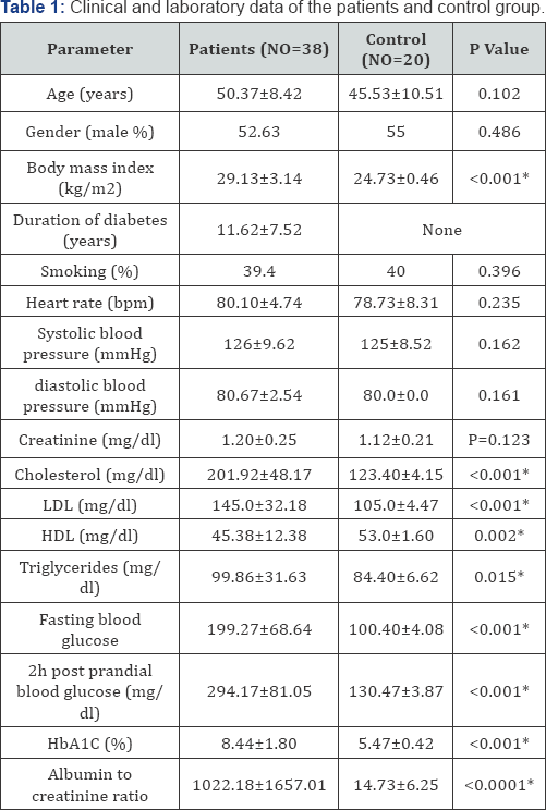
HbAIC: Glycosylated Hemoglobin; HDL: High Density Lipoprotein; LDL: Low Density Lipoproteins
The study included 38 type II DM patients aged (50.37±8.42) and 52.63% were males, 20 control subjects aged 45.53±10.51 and 55% were males with none significant difference between patient and control regarding age and gender, the mean diabetes duration was 11.62±7.52 years in patient group, regarding the laboratory data is mentioned in (Table 1).
Routine echocardiography results
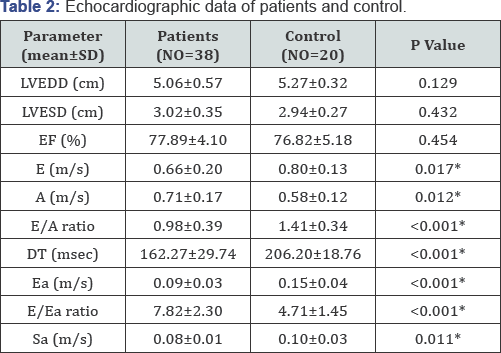
There was no significant difference between patients and control regarding LVEDD, LVESD, EF, however the E, DT, Ea, E/A, Sa was significantly lower in patients than in control, the A, E/Ea was significantly higher in patients than in control group (Table 2).
Strain and SR results
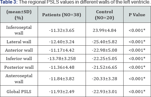
PSLS: Peak Systolic Longitudinal Strain
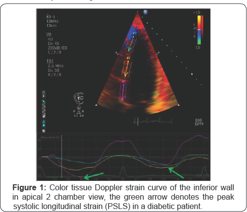
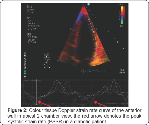

(PSSR) in a diabetic patient.
Regarding the strain results, analysis of a total 608LV segments in patients and 320LV segment in control group revealed that the regional PSLS of all walls of the LV as well as the global PSLS were significantly lower in patients than in control group (Table 3 and Figure 1). Regarding the SR results, the regional PSSR of all walls of the LV and the global PSSR were significantly lower in patients than in control group (Table 4) and (Figure 2). Correlation analysis revealed that the PSLS and PSSR was significantly negatively correlated to the duration of DM for PSLS (r=-0.705, P<0.001), and for the PSSR(r=-0.532, p=0.002) and regarding correlations with HbA1c there was also a significant negative correlation with both PSLS and PSSR (r=-0.548, p=0.002), (r=-0.532, p=0.03) respectively. By ROC curve analysis we found that a global PSLS cutoff <-15.3% can predict subclinical dysfunction in diabetic patients (AUC=0.998, sensitivity=96.67%, specificity=100%, PPV=100%, NPP=93.75 and accuracy=97.78%).
Discussion
Echocardiographic strain and strain-rate imaging is a promising tool for the evaluation of myocardial function and in our study, we evaluated regional and global LV myocardial functions by strain and SR echocardiography in asymptomatic patients with type 2DM as a method of early detection of any subclinical myocardial dysfunction and we found a subclinical regional and global LV systolic dysfunction in diabetic patients which was significantly correlated to diabetes duration and HbA1c level.
Mechanisms of decreased longitudinal myocardial function in diabetics
Longitudinal dysfunction was demonstrated in many studies using TDI derived strain and strain rate, longitudinal alteration is independently associated with diabetes mellitus, regardless of LV hypertrophy or other conventional risk factors [17].
The long term hyperglycaemia could be part of the cause, since hyperglycaemia is associated with impaired endothelium dependent relaxation caused by abnormal glycosylation end products and overproduction of free radicals [18]. Lack of this vasodilatative capacity will compromise myocardial performance [19]. Neurohumeral activation is known to influence myocardial function. Pronounced myocardial conversion of angiotensin I into angiotensin II, which is shown in DM patients, has the potential to induce increased myocardial fibrosis, in the interstitial tissue, augmented necrosis and apoptosis of the myocytes will end in loss of contractile function [20,21]. This can be aggravated by microangiopathy in the myocardial microcirculation, causing myocardial malperfusion that will increase diastolic filling pressures and enhance the extravascular component of coronary resistance. Such mechanisms can be demonstrated in the diabetic heart even in the absence of hypertension or coronary artery disease. The longitudinal subendocardial layers of the left ventricle are also susceptible to macroangiopathy in the epicardial arteries [22,23].
Comparison with other studies
Several authors demonstrated that longitudinal strain decrease was correlated with diabetes imbalance (glycosylated hemoglobin), and diabetic duration, and they were in agreement to our results. Hiromi Nakai et al. [24], Studied 55 patients with type 2DM, all patients were asymptomatic and had normal LVEF with no regional wall motion abnormalities on 2D echocardiography, they found that Regional systolic longitudinal S significantly decreased in patients group than in controls in all 16 segment and global systolic longitudinal strain significantly decreased in patients than in control group (-17.6±2.6% vs.- 20.8±1.8, p<0.001). Findings suggested that diabetic patient without overt heart disease demonstrated echocardiographic evidence of systolic dysfunction; this is in agreement with our study, also they found that diabetic duration was the only independent predictor for global strain reduction (p=0.0313); this goes with our result which suggested that duration of DM had a Significant effect on the reduction strain values in type 2 DM patients.
Zhi You et al. [25], studied 186 patients with normal EF and no evidence of CAD: 48 with DM only (DM group), 45 with left ventricular hypertrophy only (LVH group), 45 with both diabetes and LVH (DH group), and 48 normal controls. Peak strain and strain rate of six walls in apical four-chamber, long- axis, and two chamber views were evaluated and averaged for each patient and they found all patient groups showed reduced systolic function compared with controls, evidenced by lower peak strain (p<0.001) and strain rate (p=0.005) and concluded that diabetic patients without overt heart disease demonstrate evidence of systolic dysfunction and increased myocardial reflectivity. Arnold et al. [26] LV strain and strain rate analyses were used to detect subtle myocardial dysfunction in 47 asymptomatic type 2 DM compared to 53 healthy control and found that the diabetic patients had impaired longitudinal, but preserved circumferential and radial systolic, DM was an independent predictor for longitudinal strain, and systolic strain rate on multiple linear regression analysis (all p<0.001).
Vinereanu et al. [27] studied 35 patients with Type II diabetes with no symptoms, signs or history of heart disease, and 35 age and sex matched healthy controls, and found that patients with type II diabetes and no clinical heart disease have impaired subendocardial function of the LV at rest and peak stress, which is related to HBA1c and serum low-density lipoprotein-cholesterol.
Study limitation
We study only the left ventricular longitudinal function and we did not study the circumferential or radial myocardial function, this is mostly because tissue Doppler strain and strain rate imaging techniques is angle dependant, further study using speckle tracking echocardiography is recommended, also the small sample size is considered one of the limitations.
Conclusion
Type 2 DM causes subclinical LV systolic dysfunction which could be detected early using strain and strain rate echocardiography and this dysfunction is closely correlated to duration of diabetes and HbA1C level.
For more Open Access Journals in Juniper Publishers please
click on: https://juniperpublishers.com
For more articles in Open Access Journal of
Cardiology & Cardiovascular Therapy please click on: https://juniperpublishers.com/jocct/index.php
For more Open
Access Journals please click on: https://juniperpublishers.com
To know more about Juniper Publishers please click on: https://juniperpublishers.business.site/


Comments
Post a Comment