A Rare Case of Ebstein Anomaly in a French Mastiff Dog-Juniper Publishers
JUNIPER PUBLISHERS-OPEN ACCESS JOURNAL OF CARDIOLOGY & CARDIOVASCULAR THERAPY
Abstract
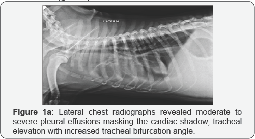
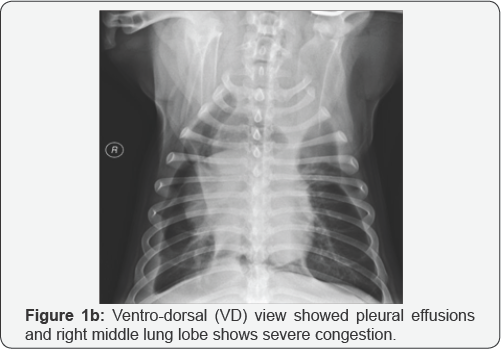
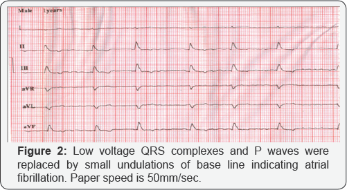
A one year old male French mastiff dog was presented with history of complete anorexia, ascites, exercise intolerance and lethargy. Chest radiographs revealed moderate to severe pleural effusions, tracheal elevation, increased tracheal bifurcation angle and pulmonary edema. Electrocardiography showed low voltage complexes and atrial fibrillation. Abdominal ultrasound revealed ascites and severe hepatomegaly. Haematobiochemical analysis revealed neutrophilic leukocytosis with mild thrombocytopenia and hypoproteinemia. Cytological and biochemical analysis of ascitic fluid indicated its modified transudate nature. Doppler echocardiography revealed tricuspid valve regurgitation with severe right atrial enlargement and there was downward displacement of tricuspid valve leaflets into the right ventricle indicating ebstein anomaly.The animal was treated with diuretics, vasodilators, ion tropic drugs for a period of four weeks. The patient died at home after four weeks of treatment and owner refused for postmortem examination.
Keywords: Ebstein anomaly; Dog; Echocardiography
Introduction
Tricuspid valve dysplasia (TVD) is a congenital deformity of tricuspid valve (TV), chordae tendineae, or papillary muscles resulting in insufficiency of tricuspid valve. The downward displacement of tricuspid valve leaflets into the right ventricle (RV) i.e. right ventricular atrialization results in a rare malformation called Ebstein's anomaly [1,2]. No doubt tricuspid valve dysplasia in dogs and cats are well mentioned in veterinary literature [3]. Ebstein anomaly is one of the rare forms of tricuspid valve dysplasia in dogs [4]. In present study, ebstein anomaly was detected in a young French mastiff dog.
Case Report
A one year old male French mastiff dog weighing 26kg was presented to Teaching Veterinary Hospital of Guru Angad Dev Veterinary & Animal Sciences University (GADVASU) Ludhiana, Punjab in Northern India with history of complete anorexia, severe ascites, groaning, exercise intolerance and lethargy for the past one month. The owner has observed that the animal tries to vomit out i.e. gagging reflux for past one month. The vaccination and deworming status was complete. There was no history of fainting or weakness, syncope or seizures, coughing and lameness. Urine and fecal output was also normal.
The physical examination revealed pale mucous membrane, strong femoral pulse, normal rectal temperature (102aF), jugular pulsation but no distension of jugular vein. Auscultation of chest revealed severe arrhythmia with grade IV systolic murmurs on right tricuspid valve area. The systolic blood pressure measured by Doppler method was 156mm of Hg (Vet-dop2, Model BF2, Vmed technology, USA)

Lateral chest radiographs revealed moderate to severe pleural effusions masking the cardiac shadow and tracheal elevation. Left bronchus was pushed up dorsally and tracheal bifurcation angle was increased and pulmonary edema in caudal lung lobe. Ventrodorsal (VD) chest radiograph showed pleural effusions and right middle lung lobe showed severe congestion (Figure 1a & 1b). The abdominal ultrasonography revealed severe ascites, extensively enlarged liver (hepatomegaly), covering almost the entire abdomen with mixed echotexture, thickened wall of gallbladder indicating cholecystitis. However spleen, kidney and urinary bladder were normal.


Electrocardiography (ECG) was performed by using Cardiart 8108 BPL six channel electrocardiographic machine with dog in right lateral recumbency. ECG examination revealed low voltage QRS complexes and P waves were replaced by small undulations of base line indicating atrial fibrillation. The R waves were 0.4mV (reference range: 2.5mV) tall and 0.06seconds (reference range: 0.05seconds) wide indicating effusions in chest cavity (Figure 2). Heart rate was 107beats per minute (reference: 70-160beats/ min.) with severe arrhythmia. Haematobiochemical analysis revealed neutrophilic leukocytosis, mild thrombocytopenia and hypoproteinemia. However, no changes were observed in rest of the parameters.
The ascitic fluid revealed numerous red blood cells, markedly degenerated neutrophils, few activated macrophages/mesothelial cells in small clumps and bacteria seen in background indicating chronic active peritonitis with focal adhesions. Biochemical analysis of ascitic fluid revealed total protein 4.4g/dl, pH-8.5, specific gravity 1.025. Total cell count of ascitic fluid was 1570/ mm3. So on the basis of cytological and biochemical analysis, the nature of ascitic fluid was modified transudate.


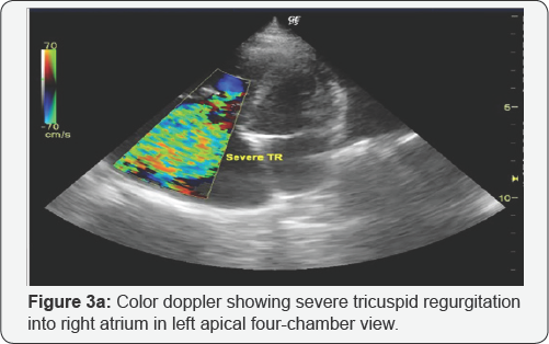
Two Dimensional, Mmode, Color flow doppler and Continuous wave doppler was performed by using GE Logiq P5 color Doppler ultrasound machine (GE healthcare, Chicago, United States) equipped with 5S transducer. Two dimensional echocardiography with Color doppler at left apical four chamber view revealed severely enlarged right atrium with tricuspid regurgitation and displacement of tricuspid valve from anatomical annular position (Figure 3a). Continuous wave Doppler echocardiography over tricuspid valve revealed a turbulent regurgitant jet with peak velocity of 2.85m/sec (Figure 3b). Based upon the modified Bernoulli equation (P=4xvelocity2), the systolic pressure gradient between right ventricle and right atrium was 32.49mm of Hg. The total length of right ventricle was 5.44cm with 1.85cm atrialized right ventricle and 5.44-1.85=3.59cm anatomical RV. The opening of tricuspid valve annulus was 5.14cm wide (Figure 3c). The characteristic sail like deformity of displaced tricuspid apparatus could be well appreciated in apical four chamber view and can be morphologically explained by tethering of septal leaflet to interventricular septum and subsequent covering of tricuspid opening by anterior tricuspid leaflet to enhance coaptation (Figure 3d). M mode echocardiography at right parasternal short axis view indicated normal left ventricular functions and paradoxical interventricular septum. Fractional shortening (FS%) and ejection fraction (EF%) were within the normal range in the present case (Figure 3e). The animal was treated with Ampicillin @25mg/kg b.wt parenterally 12hourly for 1week, furosemide (@2mg/kg, po, q12h), Enalapril (0.5mg/ kg PO every 12hr) with salt-restriction in diet. For atrial fibrillation, digoxin was given @0.008mg/kg b.wt. Four weeks after the treatment, the dog died at home and owner refused the necropsy.
Discussion
Congenital and secondary tricuspid valve abnormalities are rarely reported in dogs and cats [4-6]. The incidence of tricuspid valve dysplasia ranges from 7.0 to 7.5% out of all congenital heart defects occurring in dogs [6]. Large and pure breed dogs especially Labrador Retrievers, Boxers and German Shepherds are mostly affected [4]. Clinical signs of ascites, pleural effusions, weakness, lethargy, jugular vein pulsation and hepatomegaly suggested right sided heart failure in the present case [4,6]. Severe venous congestion in the right atrium might be responsible for occurrence of ascites and hepatomegaly [1]. Supraventricular tachycardia (SVT) is one of the common conduction abnormalities seen in human patients affected with ebstein anomaly [1]. In present case, atrial fibrillation and low voltage complexes were detected on electrocardiography without any evidence of right sided enlargement. Pleural effusions in this case might be responsible for low voltage complexes.
Echocardiography has been found be one ofthe best techniques for diagnosis of ebstein anomaly [7]. Echocardio graphically, ebstein anomaly was diagnosed on the basis of severely dilated right atrium, downward displacement of tricuspid valve leaflets into right ventricle leading to atrialization of right ventricle and severe tricuspid regurgitation [1].
A normal tricuspid valve leaflets attaches more apical than the anterior mitral leaflet in the 4-chamber view (1). However in present case tricuspid annulus was 1.85cm lower than mitral annulus and tricuspid valve (TV) was displaced toward right ventricle (RV), resulting in RV atrialization leading to Ebstein's anomaly [1]. Echocardiographic evaluation mostly shows a markedly enlarged right atrium with right ventricular volume overload. Also, the left side of heart is mostly smaller than normal [5]. Similar findings were observed in the present study. On right parasternal short axis view, right ventricular volume overload resulted in paradoxical movement of the interventricular septum and an abnormally small left ventricle as seen in the present study [5].
Initially, to reduce the volume overload in the right heart chambers, diuretic (furosemide) and an angiotensin converting enzyme inhibitor (enalapril) was used. Diuretics, ACE inhibitors and positive inotropic drugs (digoxin) are indicated for the treatment of patients affected with ebstein anomaly [1]. Diuretics and an ACE inhibitor can improve the heart function by reducing cardiac workload and fluid volume in pleural space [8]. Digoxin reduces the activation of the sympathetic nervous system and renin-angiotensin-aldosterone system [9].
So in differential diagnosis of dogs with right-sided cardiomegaly, Ebstein's anomaly should always be included [10]. Echocardiographic criteria for this anomaly are right atrial enlargement, TV regurgitation and an abnormally downward displaced TV. So owner was informed that the prognosis is poor in spite of medical therapy. The prognosis of patients with Ebstein's anomaly varies considerably depending upon the severity of the tricuspid valve leaflet displacement and subsequent tricuspid valve regurgitation, severity of right side heart failure and conduction disturbances [11].
For more Open Access Journals in Juniper Publishers please
click on: https://juniperpublishers.com
For more articles in Open Access Journal of
Cardiology & Cardiovascular Therapy please click on: https://juniperpublishers.com/jocct/index.php
For more Open
Access Journals please click on: https://juniperpublishers.com
To know more about Juniper Publishers please click on: https://juniperpublishers.business.site/


Comments
Post a Comment