Aortopulmonary Window Closure Using the Amplatzer ASD Device-Juniper Publishers
JUNIPER PUBLISHERS-OPEN ACCESS JOURNAL OF CARDIOLOGY & CARDIOVASCULAR THERAPY
Abstract
One of the relatively rare cardiac malformations
is aortopulmonary window. We report a 12-year-old boy with a mod size
aortopulmonary window who presented with an asymptomatic cardiac murmur
and in whom catheter device closure was successfully developed with an
Amplatzer ASD Cardio fix.
Introduction
One of the relatively rare cardiac malformations is
aortopulmonary window (APW), or aortopulmonary septal defect that
accounts for 0.1% to 0.6% of all cases of congenital heart disease. It
was first described by Elliotson nearly two centuries ago [1,2]. This
abnormality is characterized by an malformed communication between the
ascending aorta and the pulmonary shaft or the right pulmonary artery
[3]. The clinical manifestations of APW are not characteristic, but many
of patients have the futures of a large left to right shunt. The cases
with large APW usually have symptoms and signs of pulmonary hypertension
and congestive heart failure in the neonatal period. Severe
hypertension of pulmonary vascularity can occur in the first months of
life [4].
Closure of the aortopulmonary window defect in all
patients is essential as soon as possible. The classical treatment of
the defect is surgical closure and the median sternotomy and
cardiopulmonary bypass was recommending [2]. Current outcomes of early
repair of APW are excellent, including infants with complex associated
cardiac lesions [5]. Several case reports have been demonstrated
recently about transcatheter device occlusion of both native and
postoperative residual defects. Nowadays, several suitable devices
include Amplatzer Duct Occluder I and II, Amplatzer Septal Occluder or
ventricular septal defect closure device are used [6]. We report the use
of Amplatzer ASD Cardiofixdevice, which was originally designed for
closure of atrial septal defect, for closure of aortopulmonary window.
A 12-year-old boy presented with an asymptomatic
cardiac murmur. He has no cyanosis with full pulses, normal S1, loud S2
and a continuous murmur grade 3/6 in upper left sternal border. He had a
bounding pulse in physical exam. Chest x-ray showed mild cardiomegaly
and increased peripheral blood flow. Echocardiography confirmed a
moderate aortopulmonary window and mild pulmonary hypertension. There
were no associated cardiac anomalies. Right and left cardiac
catheterizations were performed under generalized anesthesia and
standard protocol. The course of arterial catheter passed from femoral
artery into descending aorta, ascending aorta and then into pulmonary
artery via APW respectively.
At catheterization he had mild increased pulmonary
arterial pressures (50/40 mmHg, mean = 45 mmHg) and the QP/QS was 2.5.
Aorta angiogram showed opacification of pulmonary artery from an APW.
Sizing of APW was performed by 20*40mm Numed- Boston USA sizing balloon
that showed a moderate aortopulmonary window, 7mm in size in the
ascending aorta, away from the aortic valve and coronary arteries
(Figure 1).


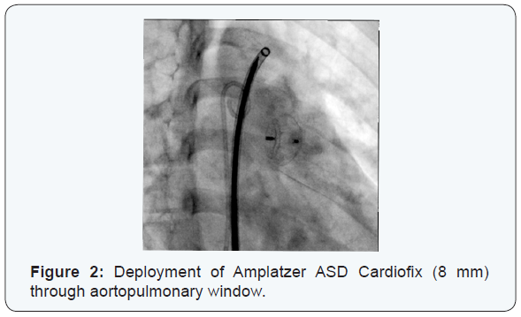
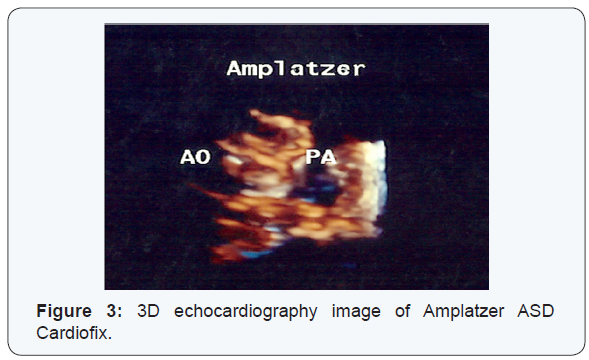
After passing delivery system through APW, Amplatzer
ASD Cardiofix (8 mm) was deployed, successfully (Figure 2
& 3). After them mean PA pressure was decreased from 45 to
24 mmHg. Echocardiography and ascending aorta angiogram
after deployment of device showed no residual APW. He was
discharged from hospital after 2 days. At follow-up 2years later,
the serial echocardiogram showed no leak through the window.
In large aortopulmonary windows, for anatomical reason,
the choice of treatment is surgery, but there are essential risks.
So, other lower risk procedures should be considered [6]. The
first using of transcatheter closure of an APW was described by
Stamato et al. They using a modified double umbrella occlude
system in a 3 years old child [7]. Tulloh and Rigby also using transcatheter umbrella closure of an APW in a child with
6-month-old by a Rash kind double umbrella system [8] Other
investigators also describe trans catheter closure of APW by
using an Amplatzer Duct Occlude device [6,9-11].
The device selection may be a preference of operator and
also related to the space available in the main pulmonary artery,
so the devices with two disks are probably better than the device
with a disk only on the aortic side. Yet, Amplatzer Duct Occluder
(ADO I) has been used to closure of APW, although the device
come out into the pulmonary artery. Alternatively, the Amplatzer
muscular ventricular septal defect occludes or ASO may be
better than ADO, or devices with similarly shaped available from
several other manufacturers [3].
Because of the device design, the device may be deployed
from the aortic side hence; a small delivery catheter is required
for it. Noonan and et.al believe that in young children, the venous
approach is preferable [6]. PremSekar, R et.al showed that the
use of the internal jugular vein can be an alternative approach to
device closure of APW in patients with occluded femoral veins
[12]. Our patient was older child who had moderate size APW
accordingly, we preferred to use the Amplatzer ASD device (size:
8 mm) and deployed it from the aortic side.
Most patients with APW develop severe clinical symptoms
such as congestive heart failure or pulmonary hypertension.
Early surgical closure or device employment is indicated as soon
as the diagnosis is established. Nonsurgical technique is possible
for closure of small aortopulmonary windows, and the range of
Amplatzer devices exhibit the several options for operators.
For more articles in Open Access Journal of
Cardiology & Cardiovascular Therapy please click on: https://juniperpublishers.com/jocct/index.php


Comments
Post a Comment