Familial Tracheooesophageal Atresia-Juniper Publishers
JUNIPER PUBLISHERS-OPEN ACCESS JOURNAL OF CARDIOLOGY & CARDIOVASCULAR THERAPY
Oesophageal Artesia most times accompanied by
tracheoesophageal fistula is not an uncommon clinical
condition. It occurs in 1 in 2,500 to 3,000 live births and there
is little evidence to support significant geographical or secular
variation in the incidence. The majority of cases of Oesophageal
Artesia are sporadic/non syndromic. Familial/syndromic cases
of oesophageal Atresia are extremely rare representing less than
1% of the total [1]. Survival is directly related to birth weight and
to the presence of a major cardiac defect [1]. Infants weighing
over 1,500 gram and having no major cardiac problem should
have a survival rate of near 100% [1,2].
We report a case of a male child born at 41 weeks gestation
after an uneventful pregnancy to a 36year old woman, through
an uncomplicated Caesarean section. The birth weight was
3,215g. There was however a family history of Oesophageal
Atresia with a distal fistula in the baby’s first cousin. The Apgar
scores were 9 at one minute and ten at five minutes. Meconium
was passed within six hours of delivery. A slight increase in
whitish oral secretions was noted. After two hours of life, the
baby developed respiratory distress despite the fact that he
had not yet been fed. The respiratory rate was 96 breaths per
minute; there was reduced intensity of breath sounds at the lung
bases and generalised rhonchi.


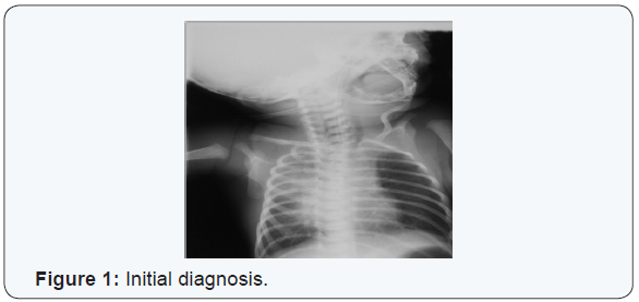
An initial diagnosis was made of aspiration pneumonitis
(Figure 1). After an initial turbulent period the baby’s clinical
condition improved. On the fourth day of life repeated attempts
to pass a nasogastric tube for feeding failed. Intraoesophageal
suction continued to yield copious aspirates. A tube containing
light barium was passed and a plain radiograph of the neck and
chest taken (Figure 2). This demonstrated a blind proximal
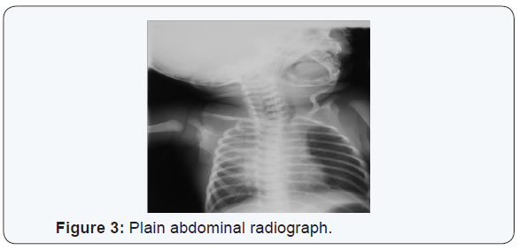
oesophageal pouch. A plain abdominal radiograph showed
abundant gastric and intestinal gas (Figure 3). Bronchoscopy
subsequently confirmed oesophageal Atresia. Clinical evaluation and an echocardiogram excluded congenital heart disease. The
baby underwent primary surgical repair on the eighth day of life
and has since recovered.
Oesophageal Atresia is a relatively common congenital
abnormality. Failure to diagnose the condition antenatally puts
the patient at risk of aspiration. At our centre in 2007 there were
3,387 live births among who were three cases of oesophageal
Atresia. There is a paucity of local data however to compare with.
The index case fits the most commonly described (86%) variety
of tracheooesophageal malformations in which there is a blind
proximal oesophageal pouch and a distal fistulous connection
with the trachea. Tracheooesophageal malformations are often
associated with other congenital abnormalities mostly cardiac
like VSD, PDA or Fallots Tetralogy and may be a component of
VATER association.
The presence of an associated defect negatively impacts on
the outcome. Our patient did not have any congenital cardiac
abnormality. One major feature of interest in the index case is
the fact that a paternal first cousin also had tracheo-oesohageal
fistula. This underlines the fact that although most cases are
sporadic, familial cases do exist [3], we wonder if this could be
a case of familial Tracheoesophageal fistula. The overall data
about Oesophageal atresia in twins reveal discordance in the
great majority with a concordance rate as low as 2.5% [4]. The
recurrence risk of the anomaly in siblings of an affected child
calculated from data from familial studies is comparable with
a recurrence risk of a multifactorial condition (1%). And the
distribution of the anomaly in first and second degree relatives
does not fit into a monogenic mode [3].
These data suggest that in most cases aetiological factors are
non genetic. No pedigree so far has been demonstrated with direct
transmission over three generations [2]. There are however,
well-defined instances where genetic factors clearly play a role
[1]. Recently three genes have been identified in humans with
a role in OA. Feingold syndrome (N-MYC), Anopthalmia – OA
(AEG) syndrome (SOX2) and CHARGE syndrome (CHDY). This is
of interest and should lead to greater insight into the aetiology of
this condition. The outcome of babies with Oesophageal atresia
and TOF has significantly improved over the last decade [4].
Prompt recognition and timely surgical intervention leads
to a good outcome in babies of weights above 1500 grammas
and the absence of cardiac and other associated anomalies. It
should be noted that fifty per cent of affected babies will have
an associated malformation and effort should be made to detect
these as soon as possible [1,2,4]. Oesophageal Atresia with
TOF remains a prime consideration in a neonate who develops feeding difficulties and respiratory distress in the first few days
of life.
Prompt recognition, appropriate clinical management to
prevent aspiration and swift referral to an appropriate tertiary
specialist centre will result in a significant improvement in rates
of morbidity and mortality. Antenatal diagnosis however allows
the delivery centre to be optimised thus improving the outcome.
Improvement in the survival of high risk cases is related to better
post operative care [6] Long term follow up is essential because
of the ongoing morbidity of the patients [5-12].
For more articles in Open Access Journal of Cardiology & Cardiovascular
Therapy please
click on:
https://juniperpublishers.com/jocct/index.php
https://juniperpublishers.com/jocct/index.php


Comments
Post a Comment