Rare Aneurysmal Bone Cyst Arising in the 3rd Rib; 5 Years Follow Up-Juniper Publishers
JUNIPER PUBLISHERS-OPEN ACCESS JOURNAL OF CARDIOLOGY & CARDIOVASCULAR THERAPY
We experienced with a relatively rare case of an
aneurysmal bone cyst (ABC) arising in the right 3rd rib. A 20-year-old
female, had experienced chest swelling on the right anterior side and
pain for 6 months. Spiral CT scan and tissue biopsy confirms aneurysmal
bone cyst of chest. Wide excision of the tumor and adjacent muscle
tissue and part of 2nd, 3rd and 4th ribs were performed with a right
antero-lateral thoracotomyincision (Sub-mammary approach). Chest wall
reconstruction was performed with prolene mesh (140 x 90 mm). In 5-year
post-operative follow up showed no recurrence.
Abbreviations: ABC: Aneurysmal Bone Cyst; CT: Computed Tomography; NIDCH: National Institute of Disease of Chest & Hospital; GA: General Anesthesia
Introduction
Aneurysmal bone cyst (ABC) is a benign tumor of the
skeletal system that rarely occurs in ribs. ABC is usually located in
the metaphysis of long bones, but very few cases involving the ribs have
been reported [1]. It is characterized by spongy, fibro-osseous,
locally destructive tissue and a multi-cystic lesion filled with blood.
Here we describe one cases of aneurysmal bone cyst in the right 3rd rib.
This disease originating in a rib was never reported in Bangladesh in
the literature.
A 20-year-old female, had experienced chest
discomfort on the right anterior side and pain for 6 months. A chest
X-ray suggested having an abnormal shadow in the left upper lung field
on the chest roentgenogram. Bilateral Mammography showed no definite
soft tissue mass or any lump within the breast with no evidence of
malignancy. Her Excision biopsy under GA with a vertical right upper
para sternal incision done on 21/01/2011. The operative finding was:
Chondroma of Ant. Chest wall.
Histopathology showed: Tissues compatible with Aneurysmal
bone cyst (ABC) with a differential diagnosis as Giant cell tumor of
bone. After a period less than a month, unfortunately the
swelling recurred with which she again consulted her surgeon with the
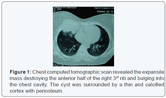
complaints of pain and swelling of the operation site.This time a spiral
CT scan of chest was done which gave an impression of large remnant of
previous mass lesion about 9x7cm mass in the upper antero-lateral chest
wall in relation to the anterior end of right 3rd rib [2].
She got admitted to NIDCH on 28/03/2011 as a known
case of post operative state of right chest wall neoplasm. She was
unmarried. She was 2nd of her two sisters and one brother. Her parents
were alive. All were in good health. She came from a middle class
family. Wide excision of the tumor and adjacent muscle tissue and part
of 2nd, 3rd and 4th ribs was performed with a right antero-lateral
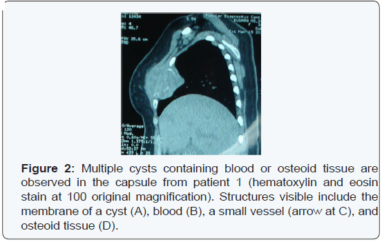
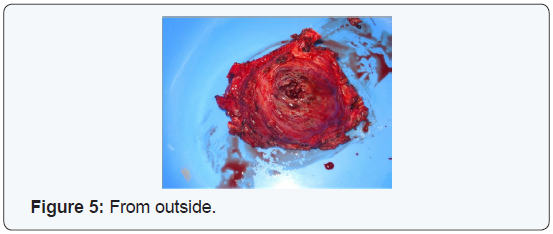
thoracotomyincision (Sub-mammary approach) on 04.04.2011 (Figures 1-5).
Chest wall reconstruction was performed with prolene mesh (140 x 90 mm).
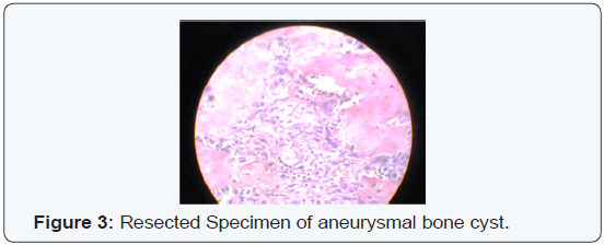
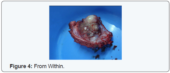
The resected specimen showed an encapsulated bony mass (75 x 60 x 35
mm) with multiple blood-filled spaces. Histopathological findings showed
multiple cysts filled with blood and fibrous trabecullae containing
osteoid tissue and multinucleated giant cells, confirming the diagnosis
of aneurysmal bone cyst. Routine post-operative follow up to 5 years
post-operatively showed no recurrence.
Aneurysmal bone cyst accounts for only 5% of all
primarybone tumors [1]. It is extremely rare in the first rib.
To those cases that have been documented in the literature to
date [3], we add a report of ABC in the 3rd rib in the Bangladeshi
population. The etiology of ABC is unclear. Some investigators
think that it may be secondary to an increased circulatory venous
pressure or trauma, causing bone absorption and blood-filled
cysts formation, thus explaining the expansile nature of ABC [1].
Others think that ABC may be secondary to other preexisting
bone diseases, giant cell tumor of bone being the most common
[4]. Symptoms of ABC in the rib are nonspecific, and it is usually
found incidentally by routine examination.
A CT scan is of great value to demonstrate the characteristic
findings of the disease. The differential diagnosis of ABC will
include Ewing sarcoma and eosinophilic granuloma, among
others [5,6]. Some have used core needle biopsy specimens to
confirm the diagnosis. We do not think this is necessary, however,
and it will increase the risk of bleeding due to the abundant
blood cysts inside the ABC. An en bloc resection with a clear
margin is still the best approach for treatment of ABC. An anterolateral
incision in sub-mammary approach allows retraction of
the mammary gland, and excellent access to the operative field.
No recurrence in our cases with the above approaches has been
observed to date.
For more articles in Open Access Journal of Cardiology & Cardiovascular
Therapy please
click on:
https://juniperpublishers.com/jocct/index.php
https://juniperpublishers.com/jocct/index.php


Comments
Post a Comment