Vascular Plug Embolization Therapy for an Unusual Pelvic Arteriovenous Malformation in a Young Adult-Juniper Publishers
JUNIPER PUBLISHERS-OPEN ACCESS JOURNAL OF CARDIOLOGY & CARDIOVASCULAR THERAPY
Abstract
The management of pelvic arteriovenous malformations
(AVMs) remains challenging due to extensive muscle plane involvement,
higher probability of incomplete surgical resection and high recurrence
rate. This report describes the case of a 24-year-old male with AVM in
the left gluteal region. Vascular surgery would have involved widespread
muscle debulking and extensive bleeding. So embolotherapy was preferred
over surgical therapy to block the blood flow into the nidus of the
AVM. The malformation was successfully treated with no recurrence after 1
year. Peripheral AVMs demands multidisciplinary approach that
integrates surgical therapy with embolotherapy. However, embolotherapy
can be exclusively developed to improve the outcomes in unusual pelvic
AVMs with very low morbidity and no recurrence.
Abbrevations: AVMs: Arterio-Venous Malformations; AVP: Amplatzer Vascular Plugs; JR: Judkin’s Right
Introduction
Arteriovenous malformations (AVMs) are vascular
anomaly characterized by abnormal connection between arteries and veins,
bypassing the capillary system [1]. Peripheral congenital AVMs are
uncommon clinical entities that can be progressive in nature,
particularly during adolescence. Specifically, pelvic AVMs are rare
vascular pathology that remains challenging to accurately diagnose and
successfully treat [2]. Diagnostic problems include poor perception of
visceral extension and precise localization [3]. Symptoms can become
debilitating and fatal if not treated well advance [4]. The management
of AVMs remains challenging because of their unpredictable behavior and
high recurrence rate [2]. In view of high bleeding risk during surgery,
numerous embolic materials have been developed, ranging from simple
gelfoam pledgets to complex systems employing micro catheters and
detachable coils [3]. However, thorough annihilation of the nidus of an
AVM is the only potential cure. Newer embolic vascular devices like
Amplatzer Vascular Plugs (AVP) are now being investigated as an
adjunctive or alternative embolic therapy for treatment of unusual
vascular pathologies like pelvic AVMs.
A 24-year-old male patient presented with a large
(appx. double the size of right gluteus) swelling at left gluteus.
Swelling was present since birth but increased since early adolescence.
In addition to cosmetic deformity, patient also experienced difficulty
in defecation due to external compression from expanding swelling over
rectum and anal canal. Auscultatory examination reveals soft continuous
shuffling sound over swelling. With consideration of non-malignant
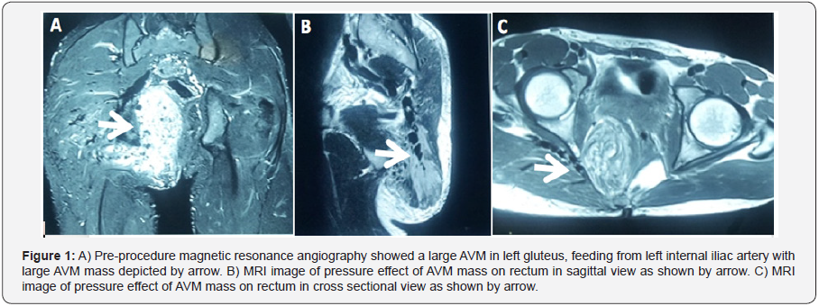
vascular pathology, patient underwent Magnetic Resonance Angiography
study which revealed large AVM originating from left internal iliac
artery (Figure 1A) with direct pressure effect on rectum and anal canal
as shown in sagittal sectional view (Figure 1B) and cross sectional view
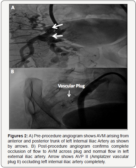
(Figure 1C). Additionally, peripheral angiogram confirmed arteriovenous
malformation arising from anterior and posterior trunk of left internal
iliac artery (Figure 2A). Vascular surgery was not considered as it
would have involved widespread muscle debulking and extensive bleeding.
Notably, high recurrence rate is also not uncommon for AVMs. So
nontraditional embolization therapy with vascular plugging was
considered.


Angiograms were reviewed for identification of the
target vessel and for other procedural details. Right SFA was
cannulated using 6F sheath. Using Judkin’s right (JR) guide
catheter (Cordis Corporation, Miami, FL) and 035 Terumo
wire (Terumo Medical Corporation, Somerset, NJ, USA), the left
internal iliac artery was selectively engaged. The 6F sheath was
exchanged with long 9F sheath. The device size was selected
based on the most restrictive diameter along the length of
the left internal iliac artery. We oversized the AVP by 30-50%,
relative to the size of the native vessel as an oversized AVP tends
to lengthen significantly. 22 mm AMPLATZER Vascular Plug II
(St. Jude Medical, Inc., Minnesota, USA) was deployed across left
internal iliac artery after carefully measuring the diameter (16
mm) and length of vessel. An adequately oversized device that
had deformed and produced a near complete occlusion of flow
suggests a tight fit and negligible chance of embolization, even in
very high flow situations [4]. Check angiogram showed no flow
across the vascular plug with normal flow in external iliac artery
(Figure 2B). Angiogram evidence of absence of flow in the AVM
demonstrated total occlusion prior to the release of AVP. The
ratio of thedevice-to-vessel diameter was 1.375.

Immediate post procedure, approximately 75% swelling
was reduced in 3 hours. Post-procedure angiogram reveals no
visible vascular mass. Any signs of periprocedural complications
including device deployment failure, device malposition,
migration or embolization, stroke, bleeding or any other
vascular complication related to the access site were closely
monitored. Patient had constipation and mild per rectal bleeding
for next 36 hours which could be due to capillary leak from
immediate decompression post-procedure. Patient was kept on
intravenous nutrition and frequent enema for 48 hours after
procedure. Patient was discharged after 72 hours and kept on
antibiotic and anti-inflammatory medication support for a week.
Upon follow up after 2 weeks, 1, 3, 6 and 12 months, patient was
asymptomatic and no signs of recurrence noted on examination.
The current management of AVMs based on the new concept
of a multidisciplinary approach can minimize the morbidity and
reduce the recurrence of the lesion. All treatments, whether
involving surgery, radiation, or drugs, have risks and side-effects.
Total excision of AVMs leads to a cure; however, total excision is
not adequate in cases of AVMs involving the joints and muscle
planes [5]. Recurrence of the AVM is common with incomplete
resection [6]. Especially, vascular malformations in pelvic region
are challenging for therapeutic management. Surgical proximal
ligation of a feeding vessel may in fact be contraindicated, because
it can make subsequent transcathetertherapy impossible [7].
There has recently been further expansion of the limited role
of embolotherapy as an adjunctive therapy for surgical resection.
This approach has even been helpful in high-risk lesions with
high-flow status [7]. Permanent occlusive agents like isobutyl
cyanoacrylate, particles of polyvinyl alcohol foam, and coils have
been used to embolize the multiple feeding vessels and even
the nidus of the AVM, whenever possible. However, recurrence
of symptoms and necessity for repeat embolization has been
reported with these devices [8]. With expanding applications in
embolotherapy, the Amplatzer series of vascular plugs are very
advantageous in the closure of a wide spectrum of abnormal
vascular communications with high technical success and low
complication rates [9]. High efficiency, lower profile, diverse
designs, reduced radiation exposure, reduced procedure
times, controlled deliverability with high precision and costeffectiveness
makes them an ideal choice for occluding various
abnormal congenital vascular communications (St. Jude Medical,
Inc. Minnesota USA).
The treatment of pelvic AVMs is a challenging topic for
vascular surgeons. Multidisciplinary treatment may offer
superior results. To minimize the complications associated
with surgery, aggressive control of blood flow is vital, and
could be pliable with a chance of cure. Our case experience of
using Amplatzer Vascular Plug has shown that transcatheter
embolization plays a substantial role in, and may be the treatment
of choice for, symptomatic pelvic vascular malformations.
Although it is demanding to outline indications, comprehensive and one-stage treatment is the ideal therapy. Extensive clinical
trials are needed to understand the definitive role of vascular
plug embolotherapy as a curative treatment of AVMs.
For more articles in Open Access Journal of
Cardiology & Cardiovascular Therapy please click on: https://juniperpublishers.com/jocct/index.php


Comments
Post a Comment