Retrieval of a Broken Peripherally Inserted Central Catheter Migrated in the Right Pulmonary Artery in a Young Girl: a Case Report-Juniper Publishers
Juniper Publishers-Journal of Cardiology

Abstract
We report a case of successful endovascular technique using a snare to retrieve a migrated, broken, peripherally inserted central catheter (PICC) in a 12-year-old girl treated with chemotherapy for lymphocytic leukemia. This patient received chemotherapy through a PICC line implanted into her left antecubital vein 9 months previously. Computed Tomography (CT) after admission showed migration of a broken PICC into the right pulmonary artery. The broken PICC was retrieved successfully through the right femoral vein using a snare. The endovascular technique is useful and should be considered initial procedure for retrieving migrated catheters.
Keywords: Peripherally inserted central catheter; Catheter migration; Retrieve; Endovascular technique
Introduction
Nowadays peripherally inserted central catheter
(PICC) is widely used in medical treatment, Catheter migration is
reported as a delayed complication which may have serious consequences
[1,2]. In cases of broken PICC migration, preferable treatments these
days are percutaneous transcatheter retrieval, open thoracotomy, being
reserved for unsuccessfully percutaneuos retrieval [3]. We report a
12-year-old girl with a 17-cm long broken PICC in the right pulmonary
artery, while the proximal end was in main pulmonary artery. The
migrated PICC was successfully retrieved by using a snare under
fluoroscopy.
Case Report
A 12-year-old girl was hospitalized for regularly
chemotherapy treatment of lymphocytic leukemia for which a PICC (Bard
access system Inc, Salt Lake City, Utah USA) had been implanted via her
left antecubital vein 9 months previously. Regular chest x-ray was
performed every 3 months in follow-up. Chest x-ray in the latest
admission showed the PICC was broken. The proximal end was shown in the
main pulmonary artery, while the distal end in the basal branch of right
pulmonary artery. The catheter stump was located in the subclavian vein
(Figure 1). CT examination confirmed migration of a broken PICC into
pulmonary artery. The distal end of the migrating PICC was in the basal
branch of the right pulmonary artery and the proximal end was adjacent
to the left wall of the main pulmonary artery (Figure 2). After initial
diagnosis, the patient received anticoagulants with warfarin (1mg/day)
according to her age and weight.
The patient was transferred to our department for
retrieval of PICC. On admission, patient was in stable condition with
normal laboratory data.
The retrieval of the broken migrating PICC was
performed 2 days after admission. Under local anesthesia, using an
ultrasound-guided percutaneous right femoral venous approach, a 6-F
catheter sheath (Terumo, Tokyo, Japan) with a 5F pig-tail catheter
(Cook, Bloomington, IN, USA) was advanced to the main pulmonary artery.
Pulmonary angiography revealed that there was no thrombus in the
pulmonary artery, and reconfirmed the position of the broken catheter
while the proximal end lay adjacent to the left wall of the main
pulmonary artery bifurcation (Figure 3). We decided to grasp the
migrating PICC at the proximal end of the catheter. With the 6-F guiding
catheter (Cordis, Miami, USA) exchange placed, a snare (Amplatz
GooseNeck, EV3 Covidien, Mansfield, MA, USA) was inserted and grasped
the proximal end of the broken PICC under fluoroscopy (Figure 4) and
successfully retrieved it through the catheter. The broken PICC segment
was 17cm in length (Figure 5). Angiography of the catheter stump in the
left upper limb showed a few small thrombus in the antecubital vein.
During the procedure, arrhythmia was present a few
seconds, and reverted to normal spontaneously. After the procedure, the
patient was treated with anticoagulant therapy: Warfarin (1mg/day) and
aspirin (50mg/day). After 2 days, the coagulation function was examined:
prothrombin time was 15.10 s and activated partial thromboplastin time
was 42.10 s. Computed tomographic angiography (CTA) on the third day
after the procedure showed no thrombus or foreign bodies in the
pulmonary vascular or subclavian vein, after which the patient was
discharged. We contact the local hospital and advice the physicians to
continue to treat the patient with Warfarin (1mg/day) for two more weeks
for safety.
Discussion
Use of central and peripheral access devices is an
integral part of modern oncology care for long-term infusion
chemotherapy. In addition, PICC may be used for total parentral
nutrition, administration of antibiotics and rehydration therapy. With
proper maintenance, they can remain in situ up to one year. Optimal
usage of PICC requires periodic (weekly) dressings and flushing.
Majority of the complications can be avoided by proper maintenance.
Common complications include phlebitis, vein thrombosis with
embolization, catheter occlusion. Catheter damage can occur with any
PICC, sometimes due to defective products but more often from improper
care. Migration of a broken catheter has been reported as a delayed
complication of PICC insertion [4,5].
Catheters can migrate at an estimated rate of
0%-3.1% within 1.5 years [6]. Migrated PICC may cause an increased
incidence of thrombosis. Cases of fatal cardiac tamponade following
migration of a broken catheter and pulmonary artery perforation have
been reported [7].
The migrated broken PICC of our patient was located
in the right branch of the pulmonary artery; it was a consequence of
migration that shifted the catheter from the insertion point into the
vein. As previously reported, this situation may have been caused by
physiological arm movement combined with the action of the
administration of fluid or frequent flushing [8,9]. The final site of
lodgment depends on the length, weight, and the material stiffness [3].
Regular chest radiography is the common choice to
ensure the safety of the inserted PICC. If it was found broken on chest
x-ray, a CT scan should be performed to find out the exact position of
the migrated PICC. Migrating PICC management includes percutaneous
transcatheter retrieval, open thoracotomy, and long-term anticoagulation
therapy. Always percutaneous endovascular technique should be attempted
first, thoracotomy being reserved for unsuccessfully cases.
CTA and/or pulmonary angiography are necessary to
exclude thrombus formation and to assist the interventional physician to
decide on which end of the catheter to grasp. In this case, the
proximal end showed on CT was adjacent to the left wall of the main
pulmonary artery, the angiography during the procedure showed it was
situated in the left wall bifurcation of the pulmonary artery and
anatomical position facilitate to grasp the proximal end with a snare.
A relative short anticoagulant therapy was
recommended in view of the young age of the patient, presumably normal
vessels and immediately lyses of small thrombus at the end of catheter.
In conclusion, the technique of percutaneous retrieval of a migrated
broken PICC is useful and should be considered by interventional
physicians for retrieving migrated catheters.
Figures and Tables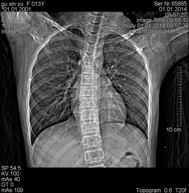 Figure 1: Chest
x-ray showed the PICC was broken. The proximal end was shown in the main
pulmonary artery (white arrow head), while the distal end deep in the
right pulmonary artery (white arrow). The catheter stump was in the
subclavian vein (white curved arrow).
Figure 1: Chest
x-ray showed the PICC was broken. The proximal end was shown in the main
pulmonary artery (white arrow head), while the distal end deep in the
right pulmonary artery (white arrow). The catheter stump was in the
subclavian vein (white curved arrow).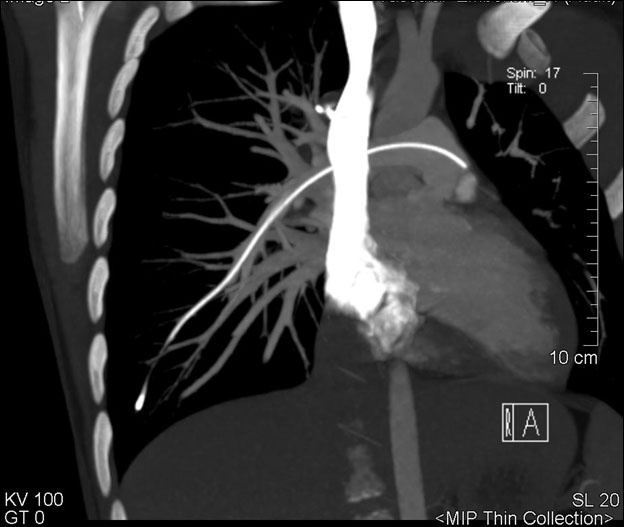 Figure 2: CTA showed
the distal end of the migrating PICC in the basal right branch of the
pulmonary artery (white arrow) and the proximal end adjacent the left
wall of the main pulmonary artery (white arrow head).
Figure 2: CTA showed
the distal end of the migrating PICC in the basal right branch of the
pulmonary artery (white arrow) and the proximal end adjacent the left
wall of the main pulmonary artery (white arrow head).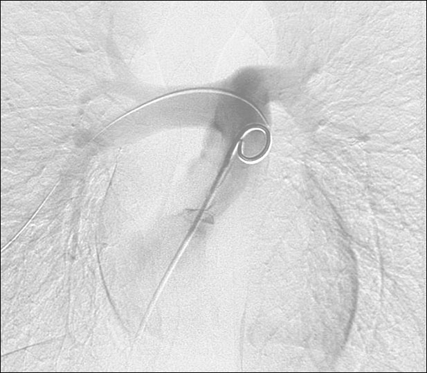 Figure 3: Pulmonary
angiography confirmed the position of proximal part of migrating
catheter in the main pulmonary trunk (black arrow head).
Figure 3: Pulmonary
angiography confirmed the position of proximal part of migrating
catheter in the main pulmonary trunk (black arrow head).
Figure 4: Under fluoroscopy the proximal end of the broken PICC was grasped by snare(black arrow).
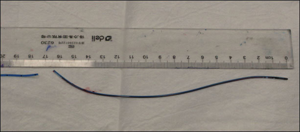 Figure 5: The broken PICC segment was 17 cm in length.
To know
more about Journal
of Cardiology Please click on :


Comments
Post a Comment